
Sending Light and Reading with Sound: What to Confirm in Photoacoustic Patch Research
Reading research that integrates laser and ultrasound receiving elements into a flexible patch.
It distinguishes the principles of operation, the scope of in vitro tests and human trials, and the limitations of external equipment connections and movements.
The Small Patch on the Skin
Can a small patch adhered to the skin visualize blood vessels within tissues? The research team led by Gao presented a photoacoustic patch in their original study published in 2022, which positions a laser diode and an ultrasound receiving element on a flexible substrate.
This article describes the research prototypes from that time and is not an introduction to any newly launched products.
What’s intriguing is not just that they reduced the size.
They created signals using the light absorption properties and constructed images by receiving those signals acoustically.
The researchers investigated the potential for hemoglobin visualization and temperature measurement.
However, the results of these two studies need to be read separately.
Below, I'll first explain the principles and results of the paper, and then add questions that the development team should consider as interpreted by MedQraft.
The visualization possibilities confirmed in humans and the diagnostic performances in patient groups are different evidences.
Light Enters and Acoustic Signals Return
The patch's laser sends short light pulses.
When the hemoglobin within the tissue absorbs the light, thermal-elastic expansion produces an acoustic signal.
The included piezoelectric element receives this signal, and the downstream system processes the signal to reconstruct a spatial image.
Describing only the transmission and reflection of typical ultrasound imaging does not fully explain this principle.
Here, light absorption is the starting point of the signal.
What wavelength of light is used and what absorbs that light connects to the image contrast.
The study focused on hemoglobin and should not be read as if they measured all the other molecules mentioned as future possibilities with the same patch.
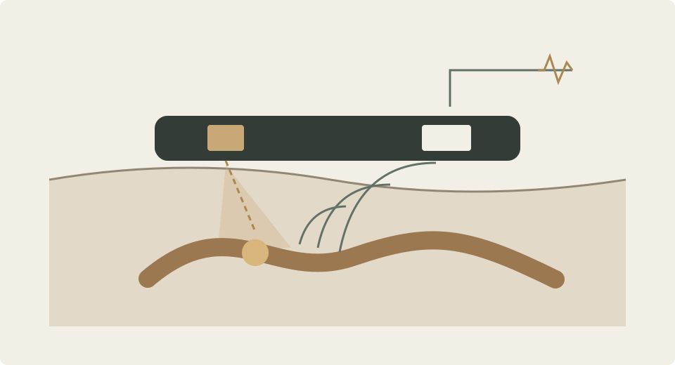
It constructs images by receiving acoustic signals generated from light absorption.
The research team wrapped the laser and receiving elements in soft materials and used a flexible connective structure.
However, not all components are equally soft.
Each element is locally solid, designed in such a way that the entire patch can follow the skin's contours.
Original Results: Design, fabrication, and working principle
Distinguishing Between Laboratory Models and Human Trials
The paper includes model tests, in vitro tests using excised tissues, and human trials together.
The expression that they used pig tissues and calf blood should not be confused with tests on living animals.
The human trial was specified in the Methods section as being conducted on a single 25-year-old male volunteer who participated under consent and institutional review.
| Test Composition | Findings Confirmed in the Study | Range to Maintain While Reading |
|---|---|---|
| Hair model in gelatin | Signal and image resolution characteristics | Distinguish between model conditions and human tissue conditions |
| Tube containing calf blood under pig tissue | Blood visualization and temperature-related signals | Interpreted as in vitro results |
| One male volunteer | Visualization of several venous regions and observation of changes | Confirmed possibilities in humans, distinct from patient group diagnostic studies |
In a comparative study with models containing blood and other liquids, photoacoustic imaging showed contrast due to differences in light absorption.
In temperature tests, the relationship between signals and temperature was examined by connecting tubes holding blood with thermocouple measurements.
This comparison provides evidence for what was confirmed in that experimental configuration.
The accuracy of distinguishing various diseases or the performance of temperature measurement for all users is not presented.
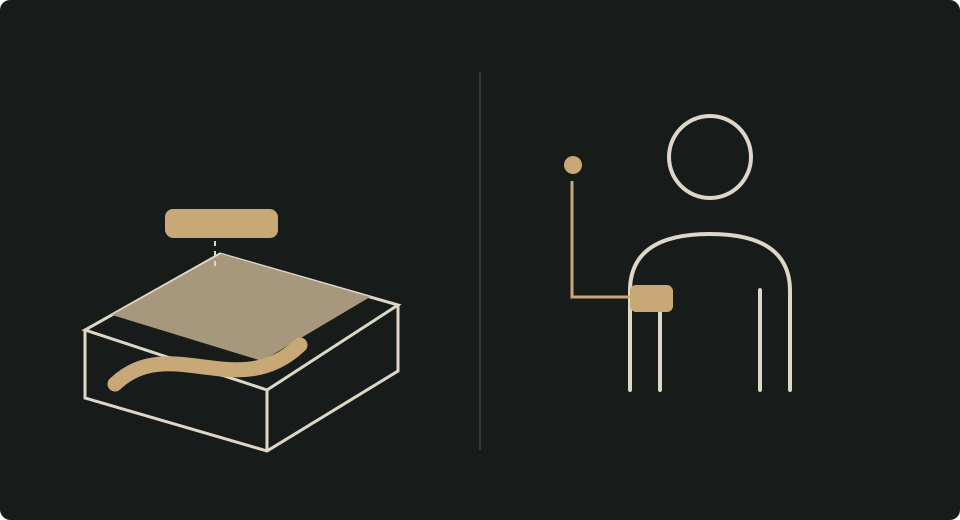
It is essential to understand the original methods where conditions were described, including placing a gelatin model between the skin and the patch to compensate for light distribution in specific vein tests.
You should check the Methods rather than guessing the entire test configuration by only looking at the patch in the picture.
Original Methods: Human experiment protocols
Numbers for Depth and Speed Come with Conditions
In the in vitro vascular model, about 2 cm thick pig tissue was visualized with a tube filled with calf blood.
In the human neck, images of internal jugular veins exceeding 1.1 cm in depth were presented.
These two values derive from different subjects and testing conditions.
It is inaccurate to combine and express, "All human blood vessels are seen at a depth of 2 cm."
In signal processing, noise was reduced through multiple averaging.
The paper’s setup achieved a 1 Hz detection frame rate with a pulse repetition of 3 kHz and 3,000 averages.
At this time, translating 1 Hz to mean that the final three-dimensional image completes automatically each second would be inaccurate.
The Methods separate the display and storage of cross-sectional images from the subsequent 3D transformation performed in dedicated software.
MedQraft's interpretation is straightforward.
When transferring performance metrics, you must include definitions of the subject, input conditions, processing stages, and output.
The term 'real-time' can refer to different aspects among signal collection, cross-sectional reconstruction, or final 3D output.
Original Results and Methods: System setup and data collection
Remaining Development Tasks
The research team described artifacts in imaging due to movement, stating that they minimized these effects by keeping the arm still during measurements.
The strong pulsations from the carotid artery in the neck affected signal averaging, which was reported as not visible in the photoacoustic images.
You should not directly generalize the results obtained from veins to arteries or moving everyday environments.
Temperature measurement also remains a separate issue.
The original text discusses the calibration relationship necessary to obtain absolute temperatures and additional tasks for applying this to humans.
Factors such as blood volume can also influence signals, so the results of in vitro temperature tests do not change to imply that deep body temperature measurements in humans are complete.
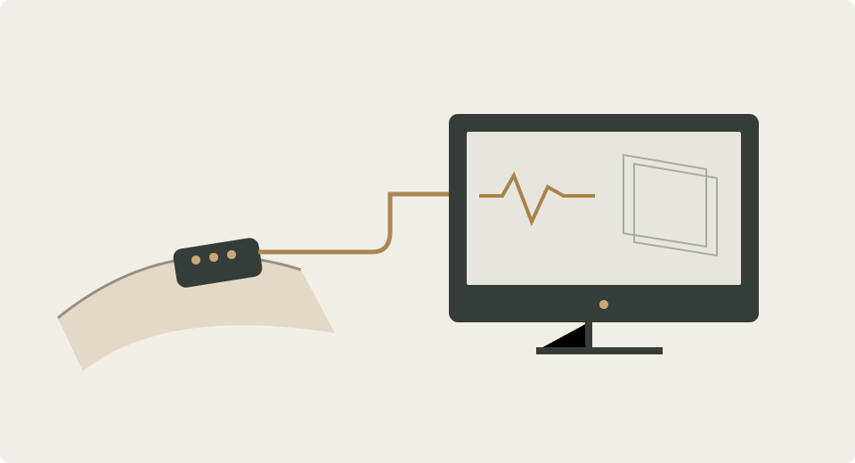
The patch was connected to external equipment via wires to collect and process signals.
Thus, saying there is a wearable sensor part is different from being a fully independent wireless system.
The researchers also suggested miniaturization and integration of the control electronics as future tasks.
Bring to a Virtual Development Meeting
Next are a hypothetical development team reviewing similar wearable imaging sensors.
This is not a real customer case or the development plan of the researchers, but an example of summarizing requirements and evaluation questions after reading the paper.
If the team's first statement is, "The vessels must be visible like in the paper," the trial goal becomes unclear.
First, you must determine which part will be observed, by whom, in what position, and how much movement will occur.
Then you can find what differences exist between the conditions of the paper and the planned use conditions.
| Hypothetical Usage Plan | Points of Contrast with the Paper | Next Questions Proposed by MedQraft |
|---|---|---|
| Observed in a moving state | Measured under stopped conditions | What are the evaluation conditions for movement and contact changes? |
| Used across different body shapes and attachment locations | Confirmation scope from one human subject | How will diversity among subjects be reviewed? |
| Portable complete system | Wired downstream equipment included | What is included in power, processing, and connection? |
| Continuous display of final 3D images | Distinction between cross-sectional display and post-processing | What are the definitions for necessary outputs and delays? |
For example, if the goal is to observe a specific area of a sitting user, there’s no need to immediately include walking scenarios into the same requirements.
On the other hand, if the actual use involves movement, it would not suffice to close out that requirement with only stationary tests.
The role of a development meeting is to narrow application scopes or define necessary evaluations backed with evidence.
Questions to Confirm When Reading the Next Paper
- What signals does the prototype create, and what does it detect?
- From what sources were the results obtained: model, excised tissues, or living subjects?
- If human subject results, how many individuals were involved, and what were the conditions?
- What comparative equipment or baseline measurements were used to confirm which questions?
- Are there depth, position, and processing conditions associated with performance metrics?
- Is equipment needed for power, collection, and processing apart from the wearable part?
- Are the researchers' prospects and the tests they actually performed separated?
These questions are not a statutory list of examinations but a proposed approach by MedQraft for reviewing papers.
This study demonstrates the potential for signal principles and sensor integration, but a single paper cannot confirm approval status or the current availability of commercial products.
To examine differences across tests further, you can read Conditions to Check Before Comparing Performance Test Results and to compare with other wearable studies, you can check Development Tasks to Read from Bladder Volume Ultrasound Research.
Original Article and Confirmation Records
Gao, X., Chen, X., Hu, H.
et al.
A photoacoustic patch for three-dimensional imaging of hemoglobin and core temperature.
Nature Communications 13, 7757 (2022).
Published on December 15, 2022.
DOI: 10.1038/s41467-022-35455-3.
Full Journal Text, PMC Full Text, PubMed Citation.
This is a peer-reviewed original article, and you can check the review information in the journal's peer review section.
No corrections or retraction notices associated with this article were found on the published journal, PMC page, or DOI search as confirmed on September 25, 2026.
The introduction, results, discussion, and methods of the text have been reviewed, though this does not mean that all supplementary materials were checked separately.
The paper is published under CC BY 4.0.
Commercial use and modifications are allowed under conditions such as attribution, and third-party materials with separate rights must be verified.
This article summarizes the original text in Korean and adds practical questions.
The original figures and tables were not reproduced; the three figures in the text, along with the hypothetical cases and review tables, were created by MedQraft.
Let's talk
Apply what you have read
to your product.
Tell us your product's current stage and the questions you need to review.
Discuss your questions ↗